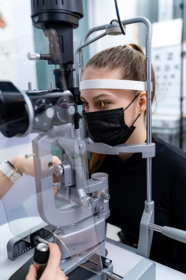
Pr Caroline FROMENT
Neuro-cognition and neuro-ophthalmology department
The service brings together the expertise relating to two specialties: the management of neurocognitive disorders and vision disorders of neurological origin.


Project
I belong to the Lyon Neuroscience Research Center (CRNL) INSERM U1028, CNRS UMRS5292, Impact Team (Integrative, Multisensory, Perception, Action and Cognition Team). My research projects concern the physiopathological understanding, clinical and functional evaluation and therapeutic management of neuro-otological or neuro-ophthalmological manifestations of neurological conditions. Within the team, I work mainly with Denis Pélisson DR and director of the team and Ruben Hermann, PHU ENT, who has just defended his science thesis which I supervised. Currently, our main projects concern oculomotor adaptation in the context of vestibular areflexia, the phenotypic characterization of CANVAS syndrome, the characterization of chronic optic neuropathies in multiple sclerosis and intracranial hypertension post dural breach closure.
Nationals collaborations
Francois Cotton, CREATIS, INSERM U1206 & CNRS UMR 5220, Lyon
- Laure Pisella, Centre de Recherche en Neurosciences de Lyon (CRNL) INSERM U1028, CNRS
UMRS5292, Equipe Trajectoire - Sandra Vukusic, Françoise Durand-Dubief, Observatoire Français de la Sclérose en Plaques,
Hôpital Neurologique Pierre Wertheimer, Bron, France - Romain Marignier, Centre de Référence des Maladies Inflammatoires Rares du Cerveau et de
la Moelle, Hôpital Neurologique Pierre Wertheimer, Bron, France - Catherine Vignal, Service de Neuro-Ophtalmologie, Fondation Rothshild, Paris
Consortium
International Clinical Consortium for NMOSD
Internationals collaborations
Aasef Shaikh, Department of Neurology, University Hospitals, Cleveland, USA Axel Petzold, The National Hospital for Neurology and Neurosurgery, University College London, London, UK Stefano Ramat, Department of Electrical, Computer and Biomedical Engineering, University of Pavia, Pavia, Italy
Publications
1. A new MRI marker of ataxia with oculomotor apraxia.
Ph Petiot, A Vighetto, F Cotton*, C Tilikete* (*Both authors contributed equally to this
paper). Eur J Radiology. 2019 Jan;110:187-192
A new MRI marker of ataxia with oculomotor apraxia — Résumé
Purpose: Evaluate the specificity and sensitivity of disappearance of susceptibility weighted imaging (SWI) dentate nuclei (DN) hypointensity in oculomotor apraxia patients (AOA).
Method: In this prospective study, 27 patients with autosomal genetic ataxia (AOA (n = 11), Friedreich ataxia and ataxia with vitamin E deficit (n = 4), and dominant genetic ataxia (n = 12)) were included along with fifteen healthy controls. MRIs were qualitatively classified for the presence or absence of DN hypointensity on FLAIR and SWI sequences. The MRIs were then quantitatively studied, with measurement of a ratio of DN over brainstem white matter signal intensity through manual delineation. The institutional review board approved this study, and written informed consent was obtained. In the cross-sectional analysis, the Mann-Whitney test was applied.
Results: Qualitatively, the eleven AOA patients presented absence of both DN SWI and FLAIR hyposignals; three dominant genetic ataxia patients had moderate SWI DN hyposignal and absent FLAIR hyposignal; the thirteen remaining subjects presented normal SWI and FLAIR DN hyposignal. Absence of DN SWI hypointensity was 100% sensitive and specific to AOA. Quantitative signal intensity ratio (mean ± standard deviation) of the AOA group (98·96 ± 5·37%) was significantly higher than in control subjects group (76.40 ± 8.34%; p < 0.001), dominant genetic ataxia group (81·15 ± 9·94%; p < 0·001), and Friedreich ataxia and ataxia with vitamin E deficit group (87·56 ± 2·78%; p < 0·02).
Conclusion: This small study shows that loss of the normal hypointensity in the dentate nucleus on both SWI and FLAIR imaging at 3 T is a highly sensitive and specific biomarker for AOA.
Keywords: Autosomal recessive cerebellar ataxia; Dentate nuclei; Genetic ataxia; Iron; Spinocerebellar ataxia; Susceptibility weighted imaging.
2. Gabapentin and memantine in treatment of acquired pendular nystagmus. Results from a
cross-over trial. »
E Nerrant, L Abouaf, F Pollet-Villard, AL Vie, S Vukusic, J Berthiller, B
Colombet, A Vighetto, C Tilikete. J Neuro-Ophthalmol 2019; 00: 1-9
Gabapentin and Memantine for Treatment of Acquired Pendular Nystagmus— Résumé
Background: The most common causes of acquired pendular nystagmus (APN) are multiple sclerosis (MS) and oculopalatal tremor (OPT), both of which result in poor visual quality of life. The objective of our study was to evaluate the effects of memantine and gabapentin treatments on visual function. We also sought to correlate visual outcomes with ocular motor measures and to describe the side effects of our treatments.
Methods: This study was single-center cross-over trial. A total of 16 patients with chronic pendular nystagmus, 10 with MS and 6 with OPT were enrolled. Visual acuity (in logarithm of the minimum angle of resolution [LogMAR]), oscillopsia amplitude and direction, eye movement recordings, and visual function questionnaires (25-Item National Eye Institute Visual Functioning Questionnaire [NEI-VFQ-25]) were performed before and during the treatments (gabapentin: 300 mg 4 times a day and memantine: 10 mg 4 times a day).
Results: A total of 29 eyes with nystagmus were evaluated. Median near monocular visual acuity improved in both treatment arms, by 0.18 LogMAR on memantine and 0.12 LogMAR on gabapentin. Distance oscillopsia improved on memantine and on gabapentin. Median near oscillopsia did not significantly change on memantine or gabapentin. Significant improvement in ocular motor parameters was observed on both treatments. Because of side effects, 18.8% of patients discontinued memantine treatment-one of them for a serious adverse event. Only 6.7% of patients discontinued gabapentin. Baseline near oscillopsia was greater among those with higher nystagmus amplitude and velocity.
Conclusions: This study demonstrated that both memantine and gabapentin reduce APN, improving functional visual outcomes. Gabapentin showed a better tolerability, suggesting that this agent should be used as a first-line agent for APN. Data from our investigation emphasize the importance of visual functional outcome evaluations in clinical trials for APN.
3. Update on Cerebellar Ataxia with Neuropathy and Bilateral Vestibular Areflexia Syndrome (CANVAS).
M Dupré, R Hermann, C Froment Tilikete. ). Cerebellum. 2021 Oct;20(5):687-700.
Update on Cerebellar Ataxia with Neuropathy and Bilateral Vestibular Areflexia Syndrome — Résumé
The syndrome of cerebellar ataxia with neuropathy and bilateral vestibular areflexia (CANVAS) has emerged progressively during the last 30 years. It was first outlined by the neurootology/neurophysiology community in the vestibular areflexic patients, through the description of patients slowly developing late-onset cerebellar ataxia and bilateral vestibulopathy. The characteristic deficit of visuo-vestibulo-ocular reflex (VVOR) due to the impaired slow stabilizing eye movements was put forward and a specific disease subtending this syndrome was suggested. The association to a peripheral sensory axonal neuropathy was described later on, with neuropathological studies demonstrating that both sensory neuropathy and vestibular areflexia were diffuse ganglionopathy. Clinical and electrophysiological criteria of CANVAS were then proposed in 2016. Besides the classical triad, frequent chronic cough, signs of dysautonomia and neurogenic pains were frequently observed. From the beginning of published cohorts, sporadic as well as familial cases were reported, the last suggestive of an autosomal recessive mode of transmission. The genetic disorder was discovered in 2019, under the form of abnormal biallelic expansion in the replication factor C subunit 1 (RFC1) in a population of late-onset ataxia. This pathological expansion was found in 100% of the familial form and 92% of sporadic ones when the triad was complete. But using the genetic criteria, the phenotype of CANVAS seems to expand, for exemple including patients with isolated neuronopathy. We propose here to review the clinical, electrophysiological, anatomical, genetic aspect of CANVAS in light of the recent discovery of the genetic aetiology, and discuss differential diagnosis, neuropathology and physiopathology.
Keywords: Bilateral vestibulopathy; Ganglionopathy; Head impulse test; Neuronopathy; RFC1; Visuo-vestibulo-ocular reflex.
4. Is the bedside head impulse test useful in emergency decision making for nonexpert
routine clinical practice? (editorial)
C Froment Tilikete. Eur J Neurol 2021 Feb 1
Is the bedside head impulse test useful in emergency decision making for nonexpert routine clinical practice? — Résumé
5. Cerebellar Signals Drive Motor Adjustments and Visual Perceptual Changes during Forward
and Backward Adaptation of Reactive Saccades.
A Cheviet, J Masselink, E Koun, R Salemme,
M Lappe, C Froment-Tilikete, D Pélisson. Cereb Cortex 2022 Jan 4;bhab455.
Cerebellar signals drive motor adjustments and visual perceptual changes during forward and backward adaptation of reactive saccades — Résumé
Saccadic adaptation ($SA$) is a cerebellar-dependent learning of motor commands ($MC$), which aims at preserving saccade accuracy. Since $SA$ alters visual localization during fixation and even more so across saccades, it could also involve changes of target and/or saccade visuospatial representations, the latter ($CDv$) resulting from a motor-to-visual transformation (forward dynamics model) of the corollary discharge of the $MC$. In the present study, we investigated if, in addition to its established role in adaptive adjustment of $MC$, the cerebellum could contribute to the adaptation-associated perceptual changes. Transfer of backward and forward adaptation to spatial perceptual performance (during ocular fixation and trans-saccadically) was assessed in eight cerebellar patients and eight healthy volunteers. In healthy participants, both types of $SA$ altered $MC$ as well as internal representations of the saccade target and of the saccadic eye displacement. In patients, adaptation-related adjustments of $MC$ and adaptation transfer to localization were strongly reduced relative to healthy participants, unraveling abnormal adaptation-related changes of target and $CDv$. Importantly, the estimated changes of $CDv$ were totally abolished following forward session but mainly preserved in backward session, suggesting that an internal model ensuring trans-saccadic localization could be located in the adaptation-related cerebellar networks or in downstream networks, respectively.
Keywords: cerebellum; corollary discharge; saccadic adaptation; trans-saccadic perception; visuo-spatial representation.
6. Scale for Ocular motor Disorders in Ataxia (SODA).
AG. Shaikh, JS Kim, C Froment, Yu Jin
Koo, …M Manto. J Neurol Sci. 2022 443:120472.
Scale for Ocular motor Disorders in Ataxia (SODA) — Résumé
Eye movements are fundamental diagnostic and progression markers of various neurological diseases, including those affecting the cerebellum. Despite the high prevalence of abnormal eye movements in patients with cerebellar disorders, the traditional rating scales do not focus on abnormal eye movements. We formed a consortium of neurologists focusing on cerebellar disorders. The consortium aimed to design and validate a novel Scale for Ocular motor Disorders in Ataxia (SODA). The primary purpose of the scale is to determine the extent of ocular motor deficits due to various phenomenologies. A higher score on the scale would suggest a broader range of eye movement deficits. The scale was designed such that it is easy to implement by non-specialized neurological care providers. The scale was not designed to measure each ocular motor dysfunction’s severity objectively. Our validation studies revealed that the scale reliably measured the extent of saccade abnormalities and nystagmus. We found a lack of correlation between the total SODA score and the total International Cooperative Ataxia Rating Scale (ICARS), Scale for Assessment and Rating of Ataxia (SARA), or Brief Ataxia Rating Scale (BARS). One explanation is that conventionally reported scales are not dedicated to eye movement disorders; and when present, the measure of ocular motor function is only one subsection of the ataxia rating scales. It is also possible that the severity of ataxias does not correlate with eye movement abnormalities. Nevertheless, the SODA met the consortium’s primary goal: to prepare a simple outcome measure that can identify ocular motor dysfunction in patients with cerebellar ataxia.
Keywords: Cerebellum; Eye movements; Gaze; Nystagmus; Rating scale; Saccades.
7. Diagnosis and classification of optic neuritis.
A Petzold, C Fraser… C Froment Tilikete, ….
GT Plant. Lancet Neurol. 2022 Dec;21(12):1120-1134.
Diagnosis and classification of optic neuritis — Résumé
There is no consensus regarding the classification of optic neuritis, and precise diagnostic criteria are not available. This reality means that the diagnosis of disorders that have optic neuritis as the first manifestation can be challenging. Accurate diagnosis of optic neuritis at presentation can facilitate the timely treatment of individuals with multiple sclerosis, neuromyelitis optica spectrum disorder, or myelin oligodendrocyte glycoprotein antibody-associated disease. Epidemiological data show that, cumulatively, optic neuritis is most frequently caused by many conditions other than multiple sclerosis. Worldwide, the cause and management of optic neuritis varies with geographical location, treatment availability, and ethnic background. We have developed diagnostic criteria for optic neuritis and a classification of optic neuritis subgroups. Our diagnostic criteria are based on clinical features that permit a diagnosis of possible optic neuritis; further paraclinical tests, utilising brain, orbital, and retinal imaging, together with antibody and other protein biomarker data, can lead to a diagnosis of definite optic neuritis. Paraclinical tests can also be applied retrospectively on stored samples and historical brain or retinal scans, which will be useful for future validation studies. Our criteria have the potential to reduce the risk of misdiagnosis, provide information on optic neuritis disease course that can guide future treatment trial design, and enable physicians to judge the likelihood of a need for long-term pharmacological management, which might differ according to optic neuritis subgroups.
8. Catch-Up Saccades in Vestibular Hypofunction: A Contribution of the Cerebellum?
RHermann, C Robert, V Lagadec, M Dupré, D Pelisson, C Froment Tilikete. Cerebellum. 2023
Jan 21.
Catch-Up Saccades in Vestibular Hypofunction: A Contribution of the Cerebellum? — Résumé
Long-term deficits of the vestibulo-ocular reflex (VOR) elicited by head rotation can be partially compensated by catch-up saccades (CuS). These saccades are initially visually guided, but their latency can greatly decrease resulting in short latency CuS (SL-CuS). It is still unclear what triggers these CuS and what are the underlying neural circuits. In this study, we aimed at evaluating the impact of cerebellar pathology on CuS by comparing their characteristics between two groups of patients with bilateral vestibular hypofunction, with or without additional cerebellar dysfunction. We recruited 12 patients with both bilateral vestibular hypofunction and cerebellar dysfunction (BVH-CD group) and 12 patients with isolated bilateral vestibular hypofunction (BVH group). Both groups were matched for age and residual VOR gain. Subjects underwent video head impulse test recording of the horizontal semicircular canals responses as well as recording of visually guided saccades in the step, gap, and overlap paradigms. Latency and gain of the different saccades were calculated. The mean age for BVH-CD and BVH was, respectively, 67.8 and 67.2 years, and the mean residual VOR gain was, respectively, 0.24 and 0.26. The mean latency of the first catch-up saccade was significantly longer for the BVH-CD group than that for the BVH group (204 ms vs 145 ms, p < 0.05). There was no significant difference in the latency of visually guided saccades between the two groups, for none of the three paradigms. The gain of covert saccades tended to be lower in the BVH-CD group than in BVH group (t test; p = 0.06). The mean gain of the 12° or 20° visually guided saccades were not different in both groups. Our results suggest that the cerebellum plays a role in the generation of compensatory SL-CuS observed in BVH patients.
Keywords: Bilateral vestibulopathy; CANVAS; Catch-up saccades; Cerebellar dysfunction; Head-impulse test; Vestibular areflexia.
9. Virtual reality set-up for studying vestibular function during head impulse test
C Desoche, G Verdelet, R Salemme, A Farnè, D Pélisson, C Froment, R Hermann. Front Neurol . 2023 Mar
29;14:1151515. doi: 10.3389/fneur.2023.1151515
Virtual reality set-up for studying vestibular function during head impulse test — Résumé
Objectives: Virtual reality (VR) offers an ecological setting and the possibility of altered visual feedback during head movements useful for vestibular research and treatment of vestibular disorders. There is however no data quantifying vestibulo-ocular reflex (VOR) during head impulse test (HIT) in VR. The main objective of this study is to assess the feasibility and performance of eye and head movement measurements of healthy subjects in a VR environment during high velocity horizontal head rotation (VR-HIT) under a normal visual feedback condition. The secondary objective is to establish the feasibility of VR-HIT recordings in the same group of normal subjects but under altered visual feedback conditions.
Design: Twelve healthy subjects underwent video HIT using both a standard setup (vHIT) and VR-HIT. In VR, eye and head positions were recorded by using, respectively, an imbedded eye tracker and an infrared motion tracker. Subjects were tested under four conditions, one reproducing normal visual feedback and three simulating an altered gain or direction of visual feedback. During these three altered conditions the movement of the visual scene relative to the head movement was decreased in amplitude by 50% (half), was nullified (freeze) or was inverted in direction (inverse).
Results: Eye and head motion recording during normal visual feedback as well as during all 3 altered conditions was successful. There was no significant difference in VOR gain in VR-HIT between normal, half, freeze and inverse conditions. In the normal condition, VOR gain was significantly but slightly (by 3%) different for VR-HIT and vHIT. Duration and amplitude of head impulses were significantly greater in VR-HIT than in vHIT. In all three altered VR-HIT conditions, covert saccades were present in approximatively one out of four trials.
Conclusion: Our VR setup allowed high quality recording of eye and head data during head impulse test under normal and altered visual feedback conditions. This setup could be used to investigate compensation mechanisms in vestibular hypofunction, to elicit adaptation of VOR in ecological settings or to allow objective evaluation of VR-based vestibular rehabilitation.
Keywords: head impulse test; vestibular function; vestibulo-ocluar reflex; virtual reality; visio-vestibular mismatch; visual feedback.
Copyright © 2023 Desoche, Verdelet, Salemme, Farne, Pélisson, Froment and Hermann.
10. Application of diagnostic criteria for optic neuritis – Author’s reply
A Petzold, Y Liu; International Consortium on Optic Neuritis (ICON). Lancet Neurol. 2023 May;22(5):376-377.
doi: 10.1016/S1474-4422(23)00110-2.
Application of diagnostic criteria for optic neuritis — Résumé
Pas de résumé

