Skull base neurosurgery
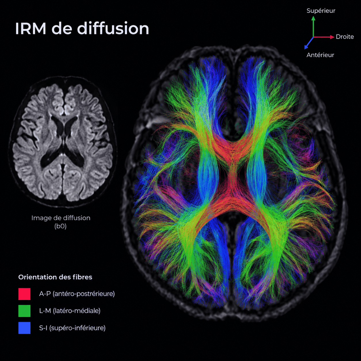
Surgery for skull base tumors remains a delicate undertaking due to the deep location of these tumors and the numerous nerves and blood vessels surrounding them. The paths of the cranial nerves, often distorted by the tumors, generally cannot be traced using conventional MRI. Professor Jacquesson and his research team employed a technique known as tractography, which is capable of mapping cranial nerve pathways in cases of complex skull base tumors. Furthermore, they demonstrated that the “pattern” displayed by the tumor on diffusion-weighted MRI could be used to predict the tumor’s histological type. Two studies published in 2025 in the *Journal of Neurosurgery* illustrate the potential of diffusion-weighted MRI to improve cranial nerve visualization and skull base tumor characterization, ultimately making surgery safer and more effective.
In an initial series of 81 skull base tumors, the tumor “pattern” observed on diffusion-weighted MRI enabled the identification of the histological type in 80.7% of cases. In a second series of 132 tumors, two methods for detecting displaced cranial nerves were compared: tractography demonstrated a significantly higher detection rate of 81.0%, compared to 53.4% for high-resolution T2-weighted MRI. However, the latter method was superior at visualizing the smallest nerves.